A Runner’s Guide to Foot Anatomy
Jun 21, 2025
 1
1
 A Runner’s Guide to Foot Anatomy www.runnerclick.com
 Our bodies are made up of fat, muscle, bones, ligaments, tendons, nerves, blood vessels, and lots of other organic matter.
As a runner, all of these things work together to cushion, support, fuel, and propel us forward.
But, our feet allow us to be able to walk, run, climb, jump, and simply be mobile.
Today we will talk about how the foot works and a runner’s guide to foot anatomy.
1. Bones
The foot is made of 26 bones. The foot skeleton provides the base structure for your muscle, tendons, and ligaments.
The way the bones in the foot are arranged allows the foot to provide balance and flexibility during movement.
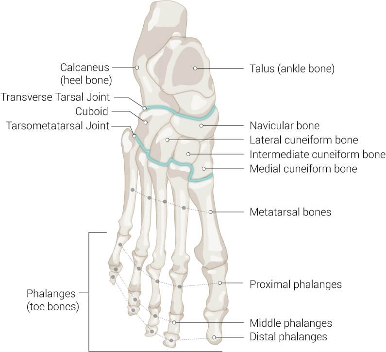
The bones of the foot begin at the base of the ankle bones, which truly are the lower end of the tibia and fibula in the leg, and end at your toes.
They consist of the following regions:
- The hindfoot
- The midfoot
- The forefoot
Hindfoot
The hindfoot is made up of talus and the calcaneus bones. The talus bones are where the tibia and fibula meet with the foot.
And the calcaneus bone is what we call our heel bone and is the largest bone in the foot.
Midfoot
The midfoot consists of the bones that create the connection between the toes and the heel.
These bones are the cuboid bone, navicular, and cuneiform bones, which are 3 separate bones.
They are more rigid, cube-like in shape, and give the foot stability.
Forefoot
The metatarsals bones and phalanges make up the rest of the foot. They give structure to the top of your foot and make up your toes.
The metatarsal bones are the connection between the midfoot and the phalanges and the upper joint connection is where you bear the most weight in your feet.
And your phalanges (toes) give you balance and assist in propulsion.
2. Joints, Ligaments, and Tendons
A joint is an area between 2 or more bones and they are often held together by ligaments.
Watertight sacks are created by ligaments and soft tissue on most of the joints in the foot to cushion and decrease friction between the joints.
In total, there are 33 joints in the foot.
Essential Joints
- Tibiotalar joint is the “ankle joint” and is where the hindfoot starts
- Transverse Tarsal joint is the joint that separates the hindfoot and the midfoot
- Tarsometatarsal joint is the joint that starts the forefoot
The curvature of these essential joints makes up the foundation of your arch. This allows for absorption of the force from your foot hitting the ground during movement.
It also assists in the flexion of your foot on different surfaces.
Ligaments and Tendons
Ligaments and tendons are very similar and are both made up of collagen. The difference is that ligaments connect bone to bone and tendons connect muscle to bone. Both of these collagenic structures provide stability.
As stated above, many of the foot ligaments make up the watertight joint capsules in the foot.
And in the foot diagram below you will see how the ligaments anchor the bones of the foot together and the bones of the ankle together.
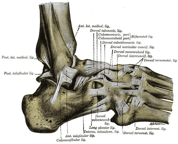
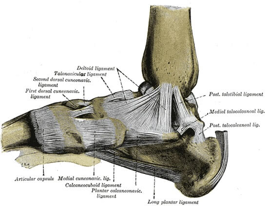
The most famous tendon that many people know about that helps us to be able to jump, run, walk, and stand upright on our feet is the Achilles tendon.
Tendons secure the muscles to bone and allow us to perform voluntary movements.
3. Muscles
The muscles of the foot are made up of lots of smaller muscles and because they are limited to the lower leg and foot only, they are called intrinsic muscles.
The 2 muscles that are on the top of the foot allow the toes to be extended. While the muscle-tendon combination on the bottom of the foot helps with flexing your toes and cushioning the bottom of the foot.
Plantar Fascia
Fascia is the collagen-containing, thin connective tissue that separates, structures, and protects organs, muscles, nerves, and bones.
It allows for the sliding of all of these systems in the body between joints and contracting movements. And fascia also helps with the synchronized movement of different systems.
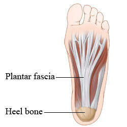
The plantar fascia (read about Plantar Fasciitis related issues), attaches from the heel bone to the base of the toes and protects the muscles in the sole of the foot.
It is also responsible for providing support to your arch in the heel-to-toe walking or running movement by sustaining the distance between the heel bone and the toes. This is called the “windlass mechanism.”
Ankle Muscles
The ankle muscle groups are arranged in compartments that are separated by strong fascia.
These compartments are the:
- Superficial posterior compartment
- Deep posterior compartment
- Anterior compartment
- Lateral compartment
They are the combination of several muscle groups that are attached to the foot by tendons to allow mobility, stability, and rolling your foot from one side to the other.
4. Nerves
Nerves allow us to feel sensations. So when you are running, you will know if you feel foot pain, have stepped on a rock, hit your big toe, got an injury such as sprains.
There are 3 nerves that enter the foot and branch off into 4 or 5 nerves; the tibial nerve, the peroneal nerve, and the saphenous nerve.
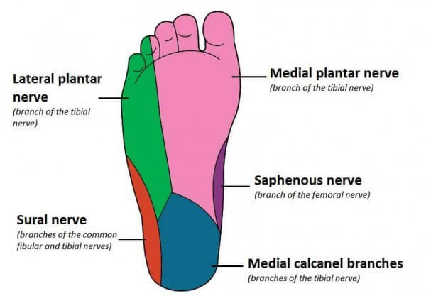
The tibial nerve branches, tibial and sural, are considered the main foot nerves and they provide sensations for the toes, the ball of the foot, and the side of the foot and control the muscles in this area.
5. Blood Vessels
There are 2 blood vessels that supply the foot with oxygen-rich blood that you will be able to feel; the posterior tibial artery and the dorsalis pedis artery.
But the main blood supply to the foot comes from the posterior tibial artery and it runs along the ankle bone on the inside of your leg.
Tie It Up
The foot is a runner’s most valued treasure. And many of our daily activities are solely dependent on our ability to be mobile.
Although the anatomy of the foot is very complex, I hope this brief overview helps you to understand your feet a little better.
Sources
- , Anatomy, Fascia, Book
- , A Patient’s Guide to Foot Anatomy, Blog
- , Anatomy of the Foot and Ankle, Orthopedic Site
- , Foot Anatomy, Online Resource
Latest Articles
 Is Running on a Treadmill Easier Than Running Outside?Runners have their own preferences, whether it is treadmill running, running outside on the road, or exploring trails. So...
 Is It OK to Use Trail Running Shoes on the Road?While trail running shoes can be used on roads, especially in situations where a runner encounters mixed terrains or pref...
 How to Fix Sore Quads After Running?Rest, ice, gentle stretching, and over-the-counter pain relievers can help soothe sore quads after running. Also, ensure ...
 10 Fruits With The Most Electrolytes to Replace Sports DrinksThese fruits are high in electrolytes such as potassium, magnesium, and calcium, essential for hydration, muscle function...